Correctly label the following parts of the femur – Correctly labeling the various parts of the femur is crucial for a comprehensive understanding of its anatomy and clinical significance. This article provides a detailed overview of the femur’s structure, including its proximal, shaft, and distal regions, highlighting their key features and functions.
Understanding the femur’s anatomy is essential for medical professionals, students, and researchers alike, as it aids in accurate diagnosis, surgical planning, and treatment of various musculoskeletal disorders affecting this bone.
Femur Anatomy

The femur, also known as the thigh bone, is the longest and strongest bone in the human body. It is located in the upper leg and plays a crucial role in weight-bearing, movement, and stability.The femur has a complex structure, consisting of several distinct regions: the proximal femur, the shaft of the femur, and the distal femur.
Each region has its own unique anatomical features and functions.
Proximal Femur
The proximal femur consists of the head, neck, greater trochanter, and lesser trochanter.The head of the femur is a smooth, spherical structure that articulates with the acetabulum of the pelvis, forming the hip joint. The neck of the femur connects the head to the shaft and is angled anteromedially.
The greater trochanter is a large, triangular prominence located laterally on the proximal femur. It serves as an attachment site for several muscles, including the gluteus medius and minimus. The lesser trochanter is a smaller prominence located medially on the proximal femur.
It provides an attachment site for the iliopsoas muscle.
Shaft of the Femur
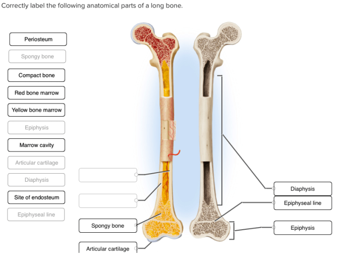
The shaft of the femur, also known as the diaphysis, is a long, cylindrical structure that extends from the proximal to the distal femur. It has a slight anterior curvature and is slightly flattened posteriorly. The shaft is composed of a thick layer of cortical bone, which surrounds a central medullary cavity.
The linea aspera is a prominent ridge that runs along the posterior surface of the shaft. It provides an attachment site for several muscles, including the vastus lateralis, vastus medialis, and adductor magnus. The nutrient foramen is a small opening located on the posterior surface of the shaft.
It allows blood vessels to enter the medullary cavity.
Distal Femur
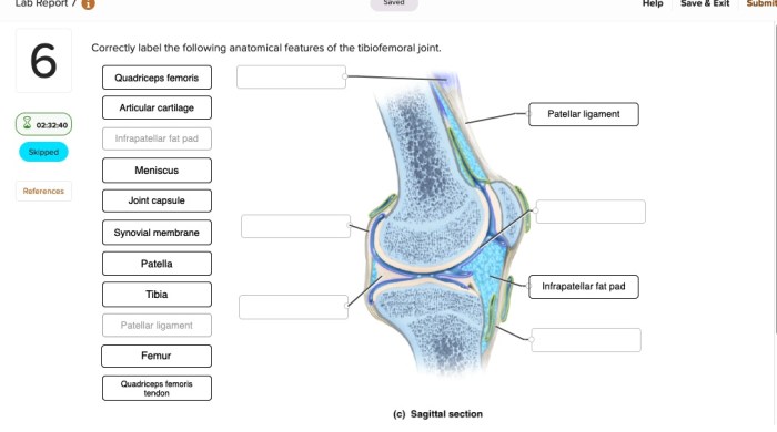
The distal femur consists of the medial and lateral condyles, the patellar surface, and the intercondylar fossa.The medial and lateral condyles are large, rounded structures that articulate with the tibia, forming the knee joint. The patellar surface is a smooth, grooved surface located on the anterior aspect of the distal femur.
It articulates with the patella, forming the patellofemoral joint. The intercondylar fossa is a deep depression located between the medial and lateral condyles. It provides an attachment site for the cruciate ligaments, which help to stabilize the knee joint.
Blood Supply and Innervation, Correctly label the following parts of the femur
The femur is supplied by several arteries, including the femoral artery, the profunda femoris artery, and the genicular arteries. The femoral artery is the main artery of the thigh and gives off the profunda femoris artery, which supplies the posterior aspect of the femur.
The genicular arteries supply the distal femur.The femur is innervated by several nerves, including the femoral nerve, the sciatic nerve, and the obturator nerve. The femoral nerve innervates the anterior and medial aspects of the femur, while the sciatic nerve innervates the posterior aspect of the femur.
The obturator nerve innervates the medial aspect of the femur.
Clinical Significance
The femur is commonly fractured due to its role in weight-bearing and its exposed position. Fractures of the femur can be classified into several types, including proximal femur fractures, shaft fractures, and distal femur fractures. Proximal femur fractures are often associated with osteoporosis and can be difficult to treat.
Shaft fractures are the most common type of femur fracture and can be caused by high-energy trauma. Distal femur fractures are often caused by falls and can involve the knee joint.The femur is also involved in several joint disorders, including osteoarthritis and rheumatoid arthritis.
Osteoarthritis is a degenerative joint disease that can affect the knee and hip joints. Rheumatoid arthritis is an autoimmune disorder that can cause inflammation and damage to the joints, including the knee and hip joints.Surgical approaches to the femur are necessary to treat fractures and joint disorders.
Common surgical approaches include the anterior approach, the posterior approach, and the lateral approach. The anterior approach is used to access the proximal femur, while the posterior approach is used to access the distal femur. The lateral approach is used to access the shaft of the femur.
Expert Answers: Correctly Label The Following Parts Of The Femur
What is the clinical significance of correctly labeling the femur?
Correctly labeling the femur is crucial for accurate diagnosis and treatment of fractures, joint disorders, and other musculoskeletal conditions involving this bone.
How does the shape of the femoral head contribute to its function?
The spherical shape of the femoral head allows for a wide range of motion at the hip joint, facilitating activities such as walking, running, and squatting.
What is the role of the linea aspera in the femur?
The linea aspera serves as an attachment site for muscles, providing stability and strength to the femur during various movements.


